Failure of a metal-on-metal hip replacement: An unusual and severe case of corrosion
Alexander Sah, MD
History
The patient is a 79-year-old female with a 5 year history of R>L hip pain. She had anterior hip replacements done elsewhere about 10 years ago. Her gradual right hip pain has worsened over the past year. She now has sharp pain in the groin and at the side of the hip. She also has a worsening limp and depends on a cane for ambulation. She has tried injections, physical therapy, and requires norco for pain control. She has failed these nonoperative treatments, and is referred for revision consultation.
Preop
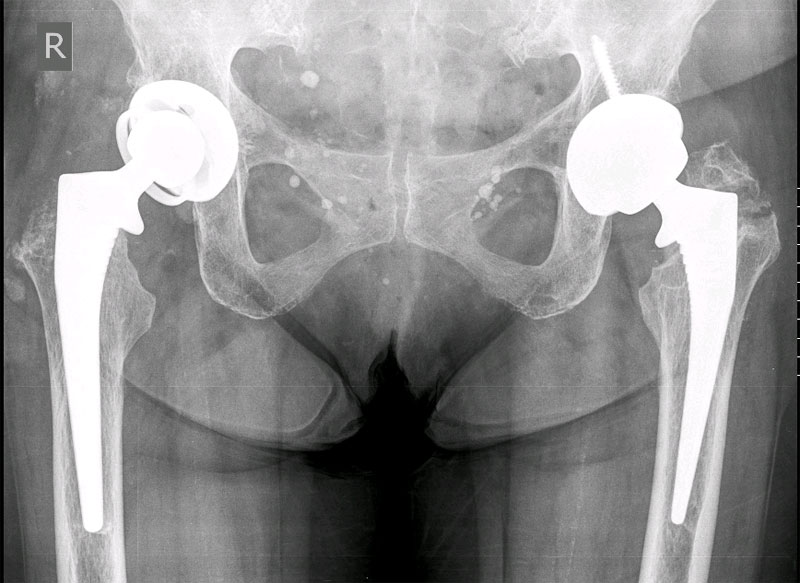
The patient has a well-healed surgical incision. She has a noticeable Trendelenburg gait. X-rays reveal a metal-on-metal hip replacement with satisfactory component alignment and fixation. Her right greater trochanter is essentially absent, from presumed osteolysis (Figure 1). Preoperative evaluation with ESR and CRP was negative for infection. Metal cobalt and chromium ion levels were significantly elevated. Of note, there is an insufficiency fracture of the greater trochanter seen on the opposite left side, also presumed to be due to corrosion and lysis.
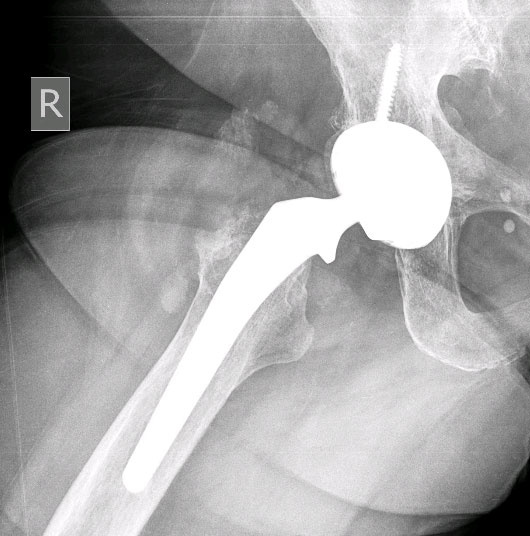
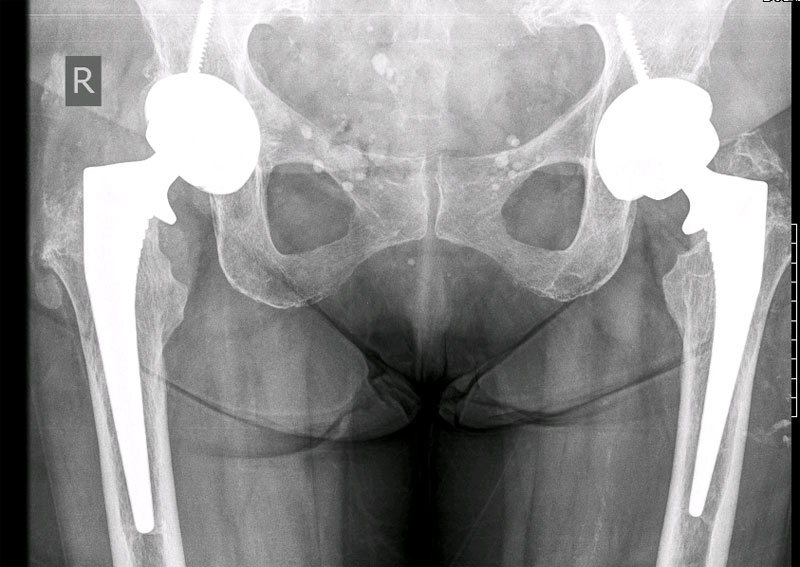
AP pelvis shows lysis of nearly the entire right greater trochanter. A fracture of the left greater trochanter is also visualized.
Surgery
Upon exposure through the fascia, a copious amount of thick discolored material was encountered (Figure 2). Material was sent for culture, which eventually returned negative for infection. Evaluation of the proximal lateral femur revealed severe osteolysis, with cortical defects laterally (Figure 3). The entire trochanter was essentially dissolved into the thick material previously removed.
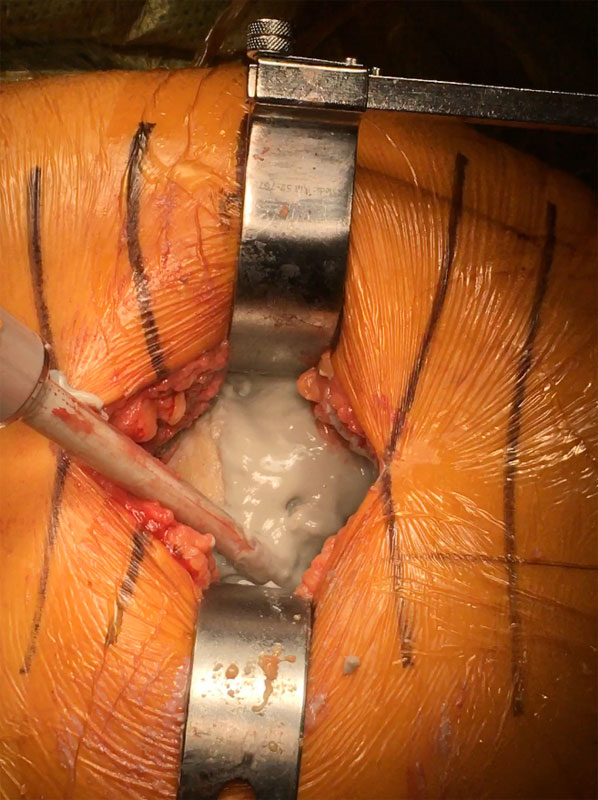
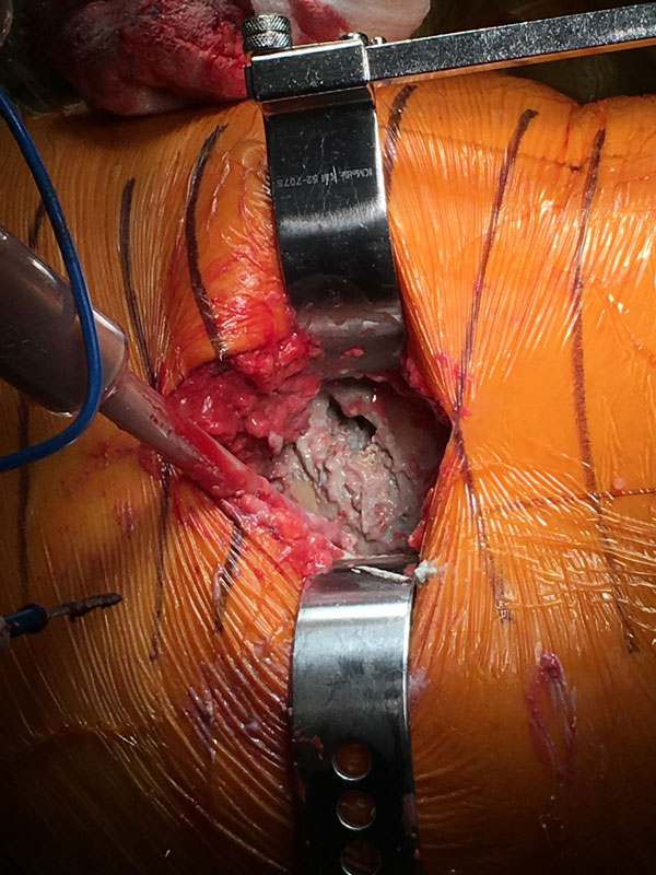
Exposure of the hip was performed and the hip dislocated. There were three areas of potential metal corrosion or metal wear. First, the backside of the metal acetabular liner was at risk due to a prominent screw head. However, there were no signs of wear at the cup-liner interface. Second, there was also no appreciable damage or wear at the liner-head articulation. Instead, at time of femoral head removal, there was significant corrosion (Figure 4).
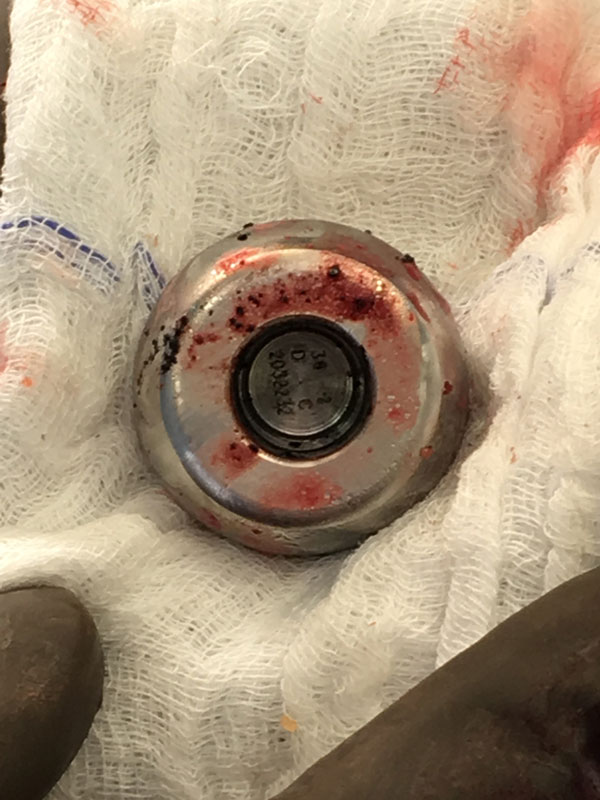
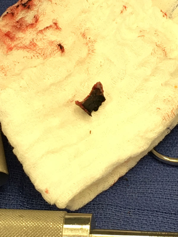
Severe corrosion discovered at the head-neck taper.
An extensive and thorough debridement was performed. The femoral stem was well fixed and retained. The taper was cleaned and a ceramic femoral head with a titanium sleeve placed. Due to the amount of abductor deficiency, and the absence of lateral proximal femur to act as host bone attachment for a potential allograft, a constrained liner was used (Figure 5).
With the extensive debridement and the amount of soft tissue damage from the corrosion process, pain management was dependent on liposomal bupivacaine injected throughout the surgical site. The exparel medication was expanded to 100cc to have more volume to disperse throughout the tissues. Revision arthroplasty procedures can benefit from having more volume of liposomal bupivacaine to optimize tissue infiltration and anesthetic overlap. A multimodal pain management protocol was also used, which allowed the patient to ambulate the day of surgery, and to be discharged home comfortably the following morning.
Postop
The patient’s pain improved almost immediately after surgery. She had an uneventful postoperative course. Her limp is mildly improved, but she understands that her abductor deficiency will persist because of the damage to her trochanter and abductors. She is relieved to have the source of her corrosion resolved, and to be able to now maximize her outcome with therapy. She is also scheduled for revision of her opposite left metal-on-metal hip replacement, which has a less severe case of corrosion.
Leave a Reply
You must be logged in to post a comment.